
#Pathresidents #GIPath #Surgpath A resection from a 2cm pedunculated polyp found on screening colonoscopy in a 60 year old man: See the thread for a poll, and some short discussion slides.

 Open web. Clear feed.
 ✓
A dynamic hub of interdisciplinary research, clinical practice, and commitment to medical education. Meet our Residents & Fellows: @BrownPathRes
#Pathresidents #GIPath #Surgpath A resection from a 2cm pedunculated polyp found on screening colonoscopy in a 60 year old man: See the thread for a poll, and some short discussion slides.
Fantastic weekend read: Oftentimes these rare entities are under recognized! Thank you as always, @LiangChengMD #brownpath #GUpath


Can’t wait to learn from the top minds at Hopkins on hot-button issues in #GUPath! Thank you, @Andres_Matoso!

Mums? Daisies? Can’t say for sure - but either way - a nice Fall-theme visual model for colon crypt histology! #Pathart #brownpath


Learn from Johns Hopkins GU pathology faculty 🔬 Contemporary Issues in GU Pathology Prostate • Bladder • Kidney • Testis • Adrenal 📅 Feb 5–6, 2027 · Pre-recorded lectures + live Q&A CME/MOC accredited Register: hopkinscme.cloud-cme.com/cou… #PathTwitter #gupath

New research could pave the way for treatments that prevent neurodegenerative diseases before they begin. As part of a multi-institution research project, Brown University professor Nicolas Fawzi and his team will study proteins at the atomic level to help researchers better understand how protein dysfunction contributes to diseases such as Alzheimer's and Parkinson's. buff.ly/khxN5YN

Plasma cells are amazing! Their appearance reflects an extraordinary commitment to antibody production: the eccentric nucleus, the abundant cytoplasm filled with rough endoplasmic reticulum, the pale Golgi hof. Features we learn early in training become more interesting as we come to understand what they represent. Some plasma cells persist for decades, producing antibodies against infections we have long forgotten. I find that remarkable. Part of our immunologic history is maintained by these cells, quietly at work in the marrow. In neoplasia, they may retain that familiar appearance while producing a protein capable of causing profound injury. A seemingly mature plasma cell can belong to a clone whose antibodies damage the kidneys, deposit in tissues, or increase blood viscosity. Its appearance alone tells us surprisingly little about the harm it can cause. (Images of plasma cell myeloma and rouleaux) #Hematopathology #Pathology #hemepath #mmsm


Flat Epithelial Atypia (FEA), left pic Focal Atypical Ductal Hyperplasia (ADH), right pic Dr. Allison. 2026 Diagnostic Pathology Update #USCAP #pathology #PathX


Words of wisdom


Gleason Pattern 4: Poorly formed glands None of these glands have lumina, this is not tangential section . Probably you could think this is grade 5. Do not call it grade 5 Across all clinical outcome groups, this morphologic pattern is NOT associated with metastatic potential. Dr. McKenney Prostatic Adenocarcinoma Grading: 2026 Update #USCAP #pathX #pathology


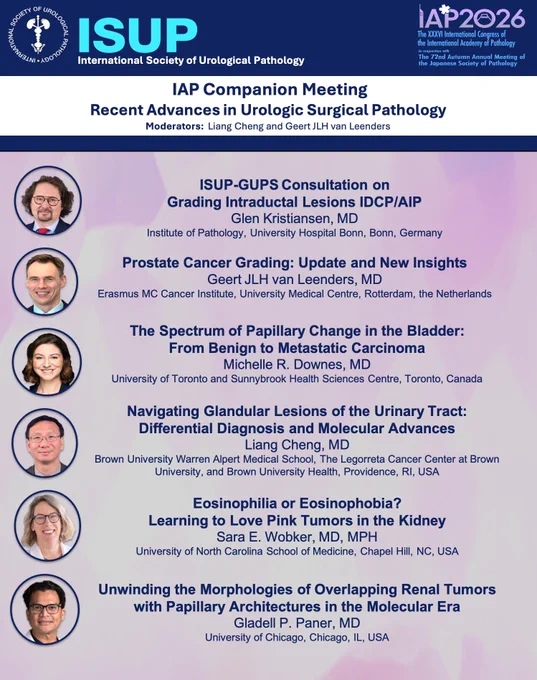
Join us at the ISUP–IAP Companion Meeting: Recent Advances in Urologic Surgical Pathology! #IAP2026 🇯🇵 🗓 Sunday, Oct 11, 2026 ⏰ 3:30–5:30 PM 📍 Palace Room (Room 9, 2nd Floor), Fukuoka International Congress Center, Fukuoka, Japan
Sincere thanks for dedication, Missy - we will miss you as you embark on your next chapter!

Can’t wait to hear the new recommendations - thank you, @LiangChengMD !


The ISUP Stockholm meeting was a tremendous success - an unforgettable gathering and an inspiring milestone for the future of precision pathology and management of GU malignancies. On September 12, 2026, 112 experts and colleagues from around the world came together at the Grand Hôtel Stockholm for a multidisciplinary expert consultation on precision biomarker testing in bladder and prostate cancer. We are living through one of the most transformative periods in the history of pathology. Cancer diagnosis is no longer defined by morphology alone. Increasingly, diagnosis, classification, prognosis, therapeutic selection, and disease monitoring are being shaped by biomarkers, molecular testing, and artificial intelligence.Our Stockholm conference embraced this transformation. Together, we examined the current and future landscape of biomarker testing in bladder and prostate cancer, including standardization of testing and reporting, specimen selection, liquid biopsy, artificial intelligence, digital pathology, and the critical unanswered questions that will shape the next generation of research and clinical practice. This meeting was not the conclusion of a process -it was the beginning of one. The conversations will continue. The evidence will evolve. Our recommendations will be refined, challenged, and strengthened. New technologies will emerge, new biomarkers will enter clinical practice, and new questions will undoubtedly arise. What will remain constant is our shared commitment to advancing science, improving standards, and ultimately providing better care for our patients. I am deeply grateful to our outstanding Working Group Chairs - Michelle Downes, Antonio Lopez-Beltran, Gladell Paner @GladellPaner , and Alessia Cimadamore, my co-Chair, Arno van Leenders - and to every member of our Working Groups for their extraordinary expertise, dedication, preparation, and countless contributions. My sincere thanks also go to every attendee who traveled to Stockholm and contributed ideas, questions, debate, and perspective. A very special thank to Katrina Collins, @katcollmd Nancy Greenland, and Alessia Cimadamore @AlessiaCim , who devoted countless hours to organizing the meeting and managing the many logistical details behind the scenes. @IntSocUropath

#GUpath: nested pattern invasive urothelial carcinoma? 😨 NOPE! just normal VonBrunn nests in the upper urothelial tract of this kidney😅


WHAT'S NEW IN NEUROENDOCRINE TUMORS (WHO 6TH EDITION 2026) Presence of any nuclear labeling with Ki67 above background is considered positive Ki67 stain is not a positive or negative stain. It is dynamically expressed throughout the cell cycle Stronger Ki67 IHC assays tend to flatten this variation Dr. Bellizzi GIPS pathcast youtube.com/live/o8IqIDiN_Jw… #pathology #PathX

