SfN is right around the corner!
Find us at Booth #1437 in Washington, D.C. from November 14-18. Whether you want to talk about your imaging, tracing, analysis setup or just catch up, we’d love to see you.
#sfn2026#neuroscience#mbfbioscience#sfn26#microscopy#3dimaging
Our multi-photon team is packing for SfN in Washington, D.C. Come see us at Booth #1437, November 14-18.
If you’re setting up new equipment, moving into high-speed imaging, or studying neural activity in freely moving animals, we can help. We’ll have:
- ScanImage and the vDAQ family for acquisition and control
- Mini2p Control Bundle for calcium imaging in freely moving mice
- SLAP2 for high-speed synaptic and voltage imaging
- vCAM a volumetric calcium imaging module based on Light Beads Microscopy that reaches up to 0.5 mm deep
Stop by to say hello, or message us to schedule a demo.
We’d love to hear about your research!
#sfn26#neuroscience#multiphotonimaging#mbfbioscience#sfn2026
MBF has reached an amazing milestone: 20,000 citations.🎉
This milestone is made possible by every researcher who has used our products and trusted MBF Bioscience with their work - thank you.
Every citation represents a scientific question someone asked, spent months at the bench investigating, and found result worth sharing. We’re so proud to have played a part, however big or small.
Your feedback over the years has made our products better and kept us focused on the work that matters. This is a celebration we share with you, our research community, and we look forward to supporting your next project.
We'd love to meet you at SfN 2026!
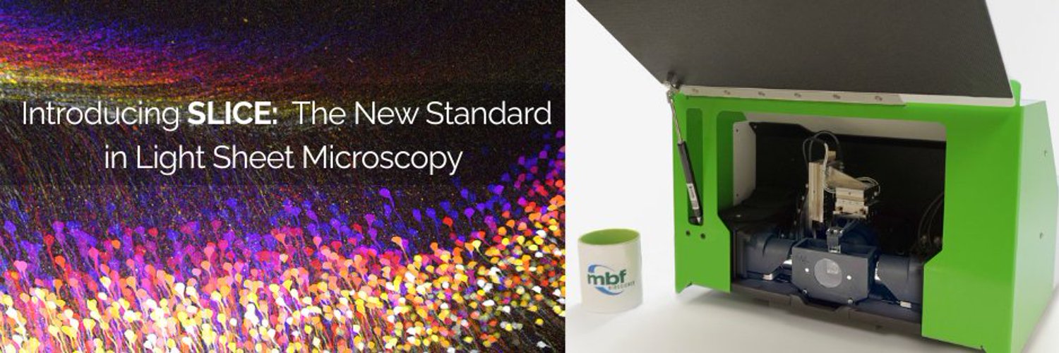
Join our team at Booth #1437 in Washington, D.C., November 14-18, for a live look at SLICE 🔬, our award-winning light sheet microscope. With its compact design and intuitive workflow, SLICE brings high-resolution 3D imaging of cleared tissue within reach for labs of all sizes.
Bring your questions and tell us about your research. To reserve a demo time, send us a message.
See you in D.C.!
📷 Image courtesy of Kevyn Jackson and Bernice Morrow, Albert Einstein College of Medicine.
#sfn2026#lightsheetmicroscopy#neuroscience#mbfbioscience#sfn26#3dimaging#sciencematters
BIG NEWS! MBF Bioscience is excited to announce a leadership transition. Effective October 1, 2026, Veronica Pessino, PhD, was appointed CEO of MBF Bioscience. Founder Jack Glaser will continue to serve the company through efforts from the Board.
Veronica joined MBF in 2025 and has served as President since January 2026. She brings a background in neuroscience research and experience leading scientific technology businesses. She will lead MBF's next chapter, with a continued focus on serving researchers through our imaging and analysis software and hardware.
Jack co-founded MBF in 1988 and under his leadership, tools like Neurolucida and Stereo Investigator became standards in quantitative neuroscience.
Thank you, Jack, and congratulations, Veronica.
As research questions get bigger and more complex, so do the experiments built to answer them. Imaging workflows increasingly span multiple instruments, real-time analysis, and decisions.
We're very excited to be developing an MHS driver for ScanImage as part of the Model Hardware Standard (MHS) research preview: a new standard for AI agents to safely operate physical equipment in scientific research and advanced manufacturing.
During the preview, we're testing how AI agents could support real-time data analysis and more connected imaging workflows, from acquisition through to the rest of the experiment, focusing on safety.
This MHS driver is being tested as part of the research preview, it is not a shipped feature. But we're glad to be testing what these new technologies could mean for microscopy.
Learn more about our work with MHS: mbfbioscience.com/mbf-biosci…
Read @AnthropicAI's announcement on the MHS research preview: anthropic.com/news/model-har…#modelhardwarestandard#scanimage#microscopy#scientificimaging#neuroscience#artificialintelligence#researchinnovation#imagingworkflows#scientificresearch#futureofscience#researchpreview
Can the rapid antidepressant-like effects of psychedelics be separated from the side effects?
New research published in Molecular Psychiatry suggests they might. Using the FP3002 Fiber Photometry System, @Yale University researchers tracked serotonin and norepinephrine activity in real time in the prefrontal cortex of mice. They found that MDMA (but not psilocybin) triggered a robust release of norepinephrine alongside serotonin. That norepinephrine appeared to suppress the classic psychedelic behavioral signature while preserving psilocybin's antidepressant-like effects.
This study highlights how real-time neurochemical recording with the FP3002 Fiber Photometry System is helping researchers uncover the complex interactions between neuromodulators, advancing our understanding of brain function and supporting the development of more targeted neuropsychiatric therapies.
Read the study: nature.com/articles/s41380-0…
.
.
#neuroscience#neurochemistry#fiberphotometry#brainscience#neuropharmacology#research#psychedelics
Heading to #fens2026 in Barcelona? Come find us at Booth #154, we'd love to show you what we've been working on.
We'll have live demos of:
🔬 SLICE™ - our award-winning light sheet microscope designed for accessible, high-performance 3D imaging
🧠 FP3002 - our fiber photometry platform for recording neural activity in freely moving animals
You'll also get a chance to explore our broader portfolio: neuron tracing, stereology, brain mapping, multiphoton imaging, and C. elegans behavior analysis.
Our team would love to hear about your research. Stop by and say hi!
📍Booth #154
📅 6–10 July
fensforum.org/
.
.
#neuroscience#lightsheetmicroscopy#fiberphotometry#brainmapping#stereology#multiphotonimaging#mbfbioscience
Featured in @naturemethods: researchers at the Max Planck Institute for Neurobiology of Behavior developed a head-mounted multiplane microscope enabling simultaneous imaging across cortical layers in freely moving mice. Using ScanImage and the high-speed vDAQ, the team recorded neuronal activity during complex behaviors.
Read more: nature.com/articles/s41592-0…#multiphotonimaging#scanimage#vdaq#mbfbioscience
Great to see this work featured by @physorg_com.
The article highlights the Nature Biotechnology publication from Dr. Raju Tomer and collaborators introducing HySIL and SCOPE, innovative optical technologies designed to make high-resolution 3D tissue imaging more accessible and scalable.
These advances have also been incorporated into the SLICE™ Light Sheet Microscope, helping bring powerful 3D imaging capabilities to researchers across neuroscience, developmental biology, cancer research, and pathology.
Read more: phys.org/news/2026-06-3d-mic…#lightsheetmicroscopy#3dimaging#neuroscience#microscopy#slicelightsheetmicroscope#lightsheetimaging